Service
Requests
We are open to cooperations with scientific partners and industry upon reasonable request. Please fill out the form below and contact us at bioprinting@i-med.ac.at.
Nanoprinting
We use a NanoOne Bio 2-photon polymerization printer to perform high‑resolution 3D micro- and nanofabrication. In combination with colored or fully transparent, biocompatible resins, we meet potential optical, biological, and technical requirements, and can produce completely customizable, CAD-design based structures. We fabricate features ranging from a few millimeters down to the sub‑micrometer scale, enabling precise prototyping and functional devices for advanced research.
Microneedles
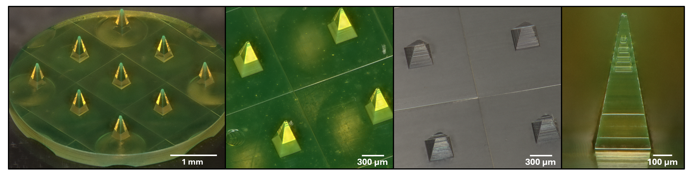
We successfully printed various needle design concepts to deliver a desired payload through the skin’s keratinocyte layer to a specific depth into the dermis:
- Hollow needles
- Porous microneedles
- Microneedle master for creating dissolvable microneedles from a PDMS mold
Microfluidic chips
We develop custom-designed microfluidic chips or modify commercially available microfluidic chips with a structural resolution down to the micrometer range.
Micro- and Nanostructure
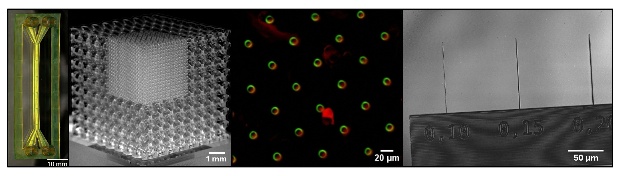
We print structures in the cellular or subcellular size range (scaffolds, pores, holes, pillars, knobs,…) to
- provide cells with a customizable three-dimensional environment in which to grow, proliferate, aggregate, or migrate
- study cell migration, cell mechanics, and cell adhesion, or to functionalize surfaces
3D-bioprinted tissue
Using 3D bioprinting, we reproduce the three-dimensional tissue architecture of normal and malignant tissue of the human body as closely as possible. Our bioprinted human tissue models represent alternatives to animal testing in the nonclinical drug development phase and are suitable test models for drug screening, toxicity testing or for investigating biological processes in a multicellular 3-dimensional context
Skin
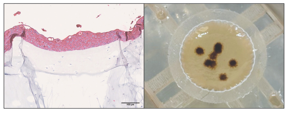
We successfully established 3D-bioprinted human skin that consists of a dermal fibroblast layer with a differentiated epidermal keratinocyte layer. The bioprinted skin models display differentiation markers analogous to normal human skin that are consistent with a fully differentiated keratinocyte layer and an intact barrier function. The bioprinted skin can be used to study the following processes:
- Preclinical drug screening
- Toxicity tests
- Skin barrier/skin penetration tests
- Skin aging
- Wound healing
Tumor models
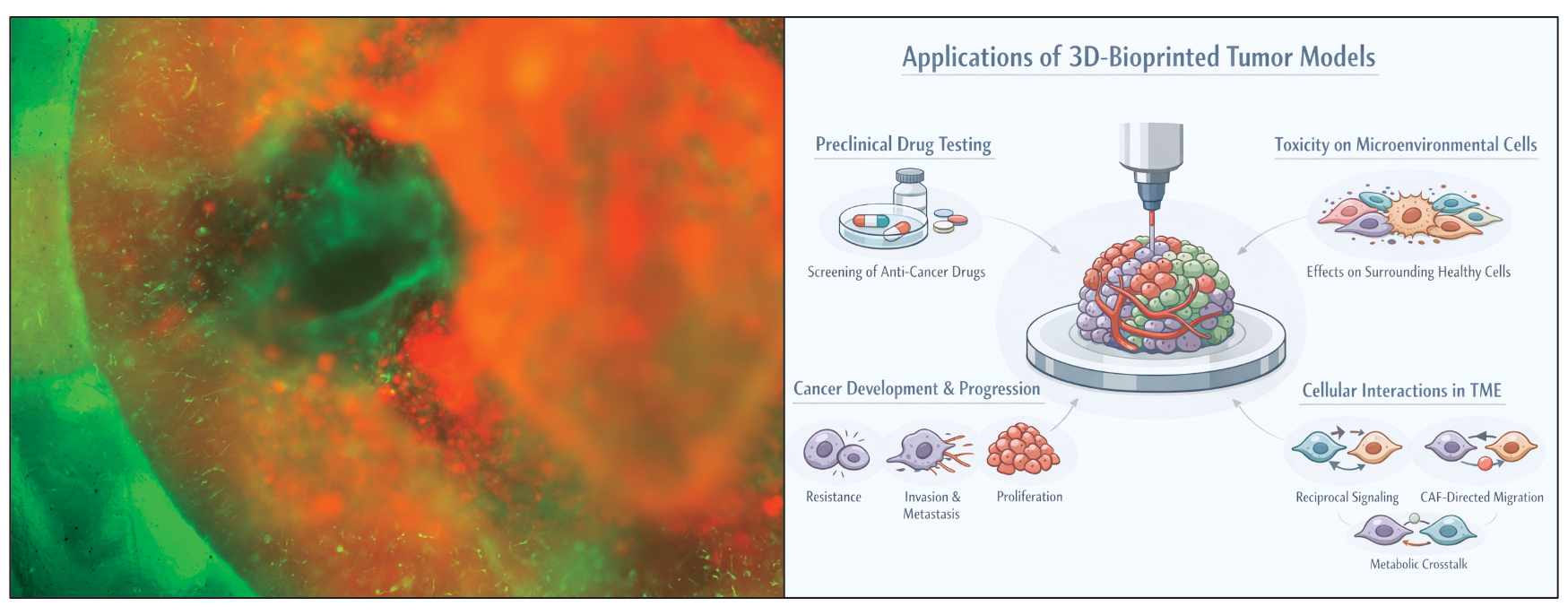
Our 3D-bioprinted tumor models combine human tumor cells with different cells of the stromal tumor microenvironment, carefully chosen to recapitulate the surroundings of the respective organ. These models allow the following analyses:
- Preclinical drug screening
- Toxicity tests on surrounding tumor microenvironment
- Biological processes of tumor development and progression (proliferation, invasion, metastasis, resistance mechanisms,…)
- Cellular interplay within the tumor microenvironment (CAF-directed migration, reciprocal crosstalk, metabolic interactions,…)